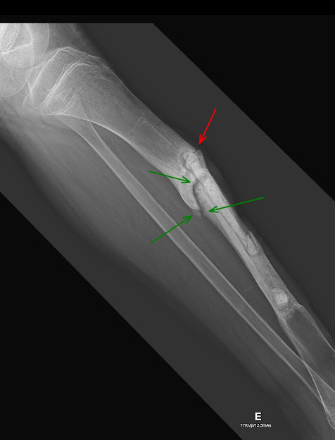
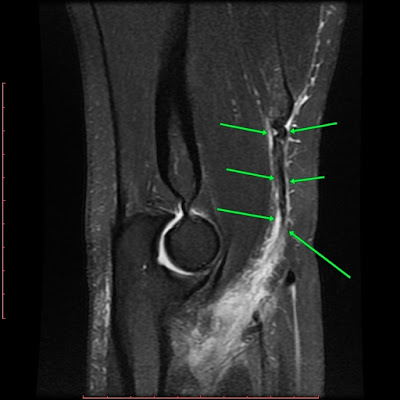
Soft tissue CHONDROMA of the Hoffa fat pad.
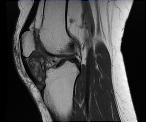
MRI. AX and SAG PD and T1. Heterogeneous expansive lobulated lesion with areas of ossification in Hoffa's fat pad.

T2.

T1 pre-contrast.

T1-post contrast (arterial phase).

T1 post-contrast (portal phase).

T1 post-contrast (transition phase).

T1 post-contrast (hepatobiliary phase). Common hepatic duct filled with with contrast (arrow)
Hemangiomas are the most common benign hepatic neoplasms, with autopsy incidence ranging from 0,4 to 20%. Typical features include high sinal intensity in T2 and specially their enhancing pattern, wich is usually pheripheral, discontinuous, nodular and progressive. Since the lesion has no hepatocellular origin, there is no uptake in the hepatobilliary phase.
Comments
Post a Comment