Posts

Transverse intermeniscal ligament rupture.

Image
MRI. AX and SAG PD FS. Partial rupture of the transverse intermeniscal ligament close to the anterior horn of the medial meniscus.  

Ramp lesion type V.

Image
MRI. AX PD FS. MRI. SAG PD FS. Double vertical tear seen in posterior horn medial meniscus.  

Fibroma of Tendon Sheath.

Image
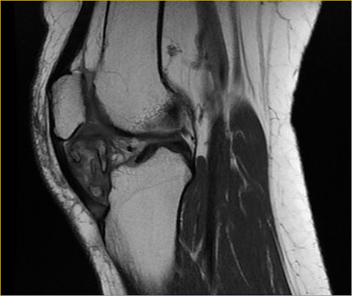
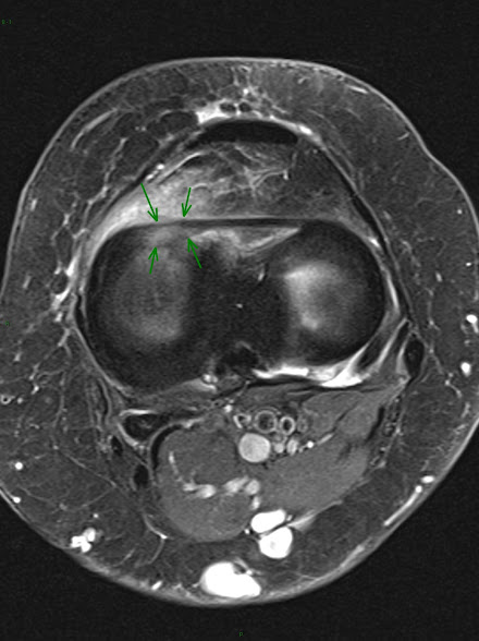
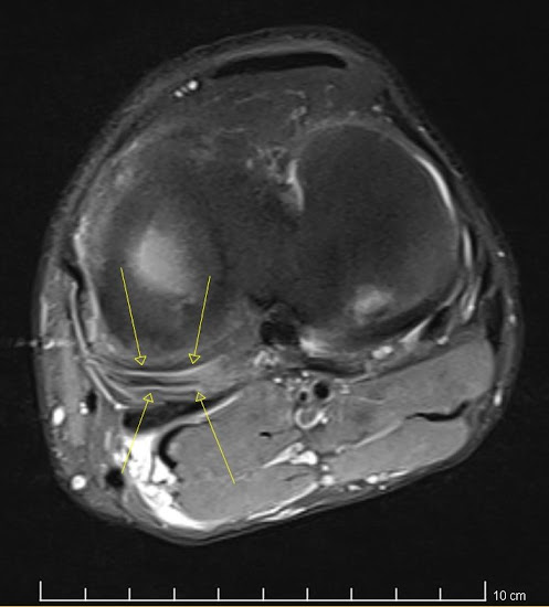
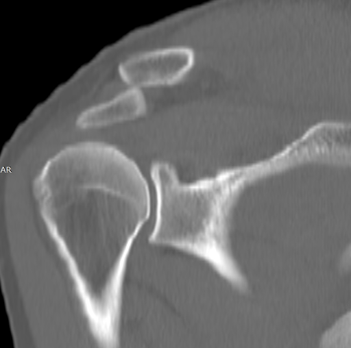
MRI. SAG T2 FS. MRI. AX T2 FS and AX T1. MRI. AX T1 FS C+.  Well-defined oval mass abutting extensor tendon with low signal intensity on T1 and T2 WI and contrast enhancement.  

Chronic Tophaceous Gout.

Image
X-ray. Foot. Multiple tophi with faint calcifications, destructive arthropaty and eccentric erosions.  

Fibular hemimelia.

Image
MRI. SAG PD. X-Ray. AP.  Type I: hypoplasia of the fibula  

Os acromiale.

Image
MRI. T1 SAG. X-ray. Neer. MRI. T2 FS. Os acromiale: mesoacromial subtype with degenerative changes.  

Salter-Harris type IV Fracture

Image
MRI. Ankle. Multiplanar fracture extends through the epiphysis, across the physis and through the metaphysis. 12 years old.  

Greenstick fracture

Image
X-ray. Incomplete fracture of radial shaft.  

Acromioclavicular Joint Dislocation Type IV

Image
CT. Complete rupture of A.C and C.C. ligaments. Clavicle displaced dorsal and posterior over acromion.  

Pellegrini-Stieda lesion

Image
X-ray. Ossified post-traumatic lesion at the medial femoral collateral ligament adjacent to medial femoral epicondyle.

Partial Lateral Patellar Facetectomy.

Image
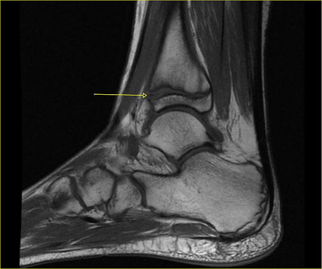
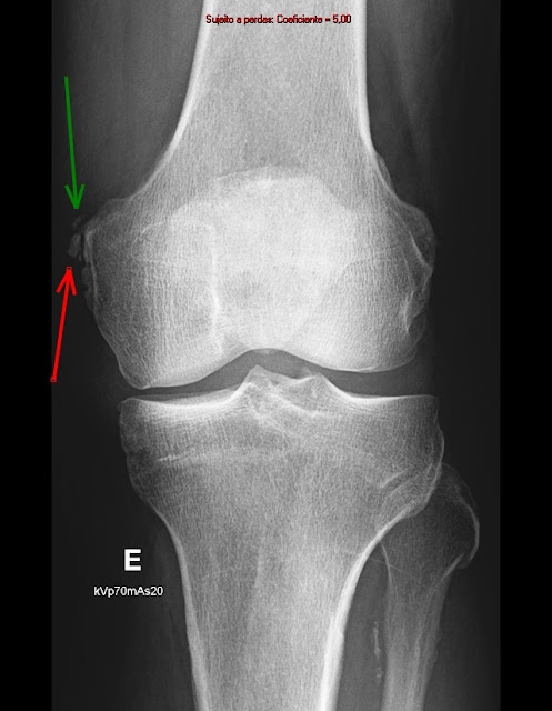
        MRI. AX PD FS.   X-ray.  Patellar Facetectomy.   X-ray.  Normal Patella.   Partial lateral patellar facetectomy for treatment of arthritis due to lateral patellar compression syndrome.

Subchondral insufficiency fracture.

Image
MRI. COR PD FS. Small fracture below the chondral surface on the weight-bearing portion of  femoral condyle with surrounding bone marrow edema.

Medial tibial plateau stress fracture.

Image
  MRI. SAG PD.