Posts

Showing posts from July, 2020

Pseudoarthrosis.

Image
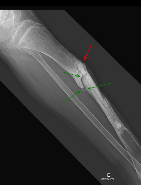
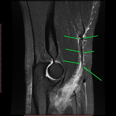
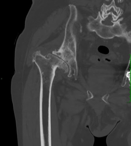
X-Ray. Postoperative control of Ewing's Sarcoma. Fracture non-union with irregular contours and bone sclerosis in the upper region of the surgical cavity in tibial shaft.

Distal biceps tendon rupture

Image
MRI. Elbow. Complete biceps tendon rupture at radial tuberosity insertion, with cranial retraction of the tendon stump and small hematoma.

Hydroxyapatite crystal deposition disease (HADD): intraosseous migration of calcification.

Image
MRI:   Rotator cuff calcifications and cystic lesion in the greater humerus tuberosity with formation of a liquid-calcium level.

Morton Neuroma

Image
T2. T1. MRI. Hypointense oval lesion in T1 and T2.

Os Hamuli Proprium

Image
MRI. Fusion failure of the hamate bone hook ossification center.

Deep vein thrombosis (DVT): fibular veins.

Image
A B MRI. A) T1. B) T2. Clinical suspicion of muscle stretching. Acute thrombus with T1-weighted hyperintense signal and surrounding soft tissue edema.

Hydroxyapatite crystal deposition disease (HADD): inflammatory / resorptive phase.

Image
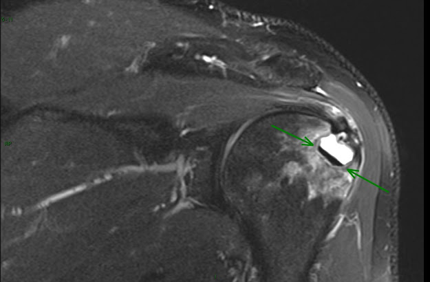
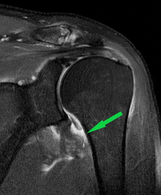
Supraspinatus tendinopathy with small partial bursal rupture and presence of large calcification extending from the critical zone to the posterior myoaponeurotic junction, with extensive swelling of surrounding soft tissues. 

Humeral avulsion of the glenohumeral ligament (HAGL)

Image
Avulsion of the inferior glenohumeral ligament (IGHL) from its humeral insertion with periarticular edema.

Developmental dysplasia of the hip (DDH): sequel in adult.

Image
CT. Dysplasia of the acetabulum, subluxation of the femoral head, secondary degenerative changes, signs of coxa magna and brevis.

Meniscus: Radial Tear.

Image
MRI. Radial tear in the posterior horn of medial meniscus.

Total Hip Arthroplasty: Wear of the Acetabular Polyethylene Liner

Image
CT. Subluxation of the hip prosthesis head, loosening of the acetabular component of the prosthesis and bone erosion in the acetabulum.

Hoffa and Prefemoral Fat Pad Impingement

Image
MRI. Hoffa and Prefemoral Fat Pad edema, usually due to continuous and repeated microtrauma.

Medial Meniscus: Grade II degenerative change. (Menisco Medial: Alteração degenerativa grau II.)

Image
MRI. Grade II:  linear area of hyperintensity, no extension to the articular surface.

Patellar Condropathy (Condropatia Patelar)

Image
MRI. Deep fissure. F issura profunda. MRI. Delamination. D elaminação.