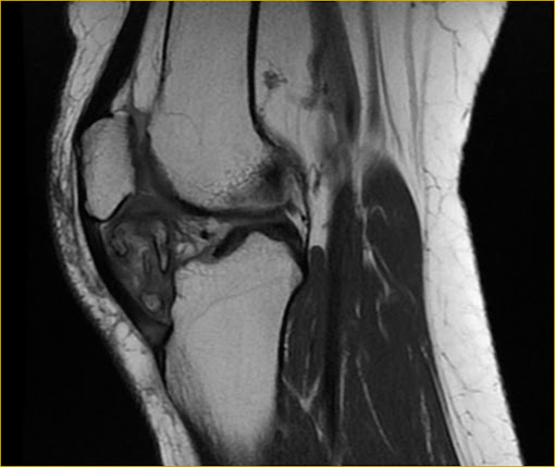
Soft tissue CHONDROMA of the Hoffa fat pad.

MRI. AX and SAG PD and T1. Heterogeneous expansive lobulated lesion with areas of ossification in Hoffa's fat pad.
Radiological cases of daily practice including magnetic resonance imaging (MRI), computed tomography (CT), positron emission tomography (PET/CT) and X-Rays, without wasting time in your routine. Click on READ MORE button to enlarge images and click on the SUBSCRIBE button to receive e-mail updates. Thank you.

WacacWstinma-1994 Rex Wright https://wakelet.com/wake/pRW6NsDW4ziAWoaGkAV6e
ReplyDeletesiecontantme
McrimverMdispge Andrew Delacuadra download
ReplyDeletegreaticirin