Radiological cases of daily practice including magnetic resonance imaging (MRI), computed tomography (CT), positron emission tomography (PET/CT) and X-Rays, without wasting time in your routine. Click on READ MORE button to enlarge images and click on the SUBSCRIBE button to receive e-mail updates. Thank you.
CT. Heterotopic Ossification. Soft tissue ovoid mass with densely calcified peripheral rim located in pre-femoral fat pad and sorrouding prominent inflamation.
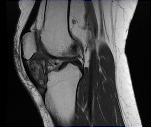
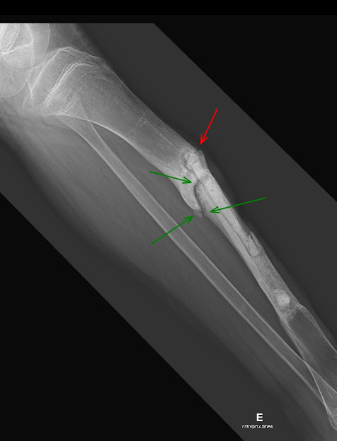
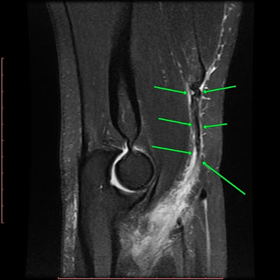
X-Ray. Postoperative control of Ewing's Sarcoma. Fracture non-union with irregular contours and bone sclerosis in the upper region of the surgical cavity in tibial shaft.


Comments
Post a Comment